We can't find the internet
Attempting to reconnect
Something went wrong
Hang in there while we get back on track

£144.99
Springer Atlas of Confocal Laser Scanning In-vivo Microscopy - Ophthalmology
biography
Price data checked 5 days ago
We'll watch every seller, every day. One email when your price arrives.
This is the most expensive it has ever been. Walk away.
£145 today · previous high £145 · all-time low £135
NEW HERE?
Amazon shows you one price. We show you all of them.
Tosheroon watches Amazon prices so you don't have to. Every product on Amazon has a price history — we make it visible. Set the price you'd actually pay, and we'll email you the second it gets there. No app, no account, one email.
WHAT'S ON THIS PAGE
when this has been cheap or pricey
where the price is heading next
all-time high & low, recent range
name your number, we'll email you
Price History & Forecast
Grey patches = out of stock. Cheaper = lower on the chart. Hover for exact prices.
Last 86 days · 86 data points (no recent data)
Price Distribution
Price distribution over 86 days • 3 price levels
Price Analysis
Most common price: £135 (59 days, 68.6%)
Price range: £135 - £145
Price levels: 3 different prices over 86 days
Description
Key Features
Confocal laser scanning microscopy technology produces in-vivo images of ocular surfaces with exceptional clarity and detail comparable to histological examination.
Comprehensive coverage of normal corneal, limbal and conjunctival anatomy with quantitative cell-density calculations for anatomical assessment.
Correlates novel optical sections of ocular surface diseases with clinical findings to support diagnostic decision-making in eye care practice.
High-magnification optical sections enable detailed examination and interpretation of corneal and conjunctival tissue structures.
Published by Springer, this atlas serves as a reference textbook for optometrists, ophthalmologists and allied health professionals specialising in ocular imaging.
Supports both diagnostic evaluation and therapeutic planning through detailed imaging principles and clinical applications in optometry.
Product Specifications
- Brand
- Springer
- Model
- biography
- Format
- hardcover
- ASIN
- 3540327053
- Domain
- Amazon UK
- Release Date
- 18 September 2006
- Listed Since
- 11 December 2006
Barcode
No barcode data available
Similar Products You Might Like

Advances in Medical and Surgical Cornea: From Diagnosis to Procedure (Essentials in Ophthalmology)
Springer
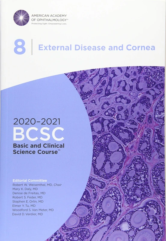
2020-2021 Basic and Clinical Science Course (BCSC), Section 08: External Disease and Cornea
American Academy of Ophthalmology

Mastering Corneal Surgery: Recent Advances and Current Techniques
Slack

Corneal Infection and Inflammation: A Colour Atlas
CRC Press

Corneal Collagen Cross Linking
Springer

Complications in Corneal Laser Surgery
Springer
2018-2019 Basic and Clinical Science Course (BCSC), Section 8: External Disease and Cornea
American Academy of Ophthalmology
Price unavailable
Corneal Regenerative Medicine: Methods and Protocols: 1014 (Methods in Molecular Biology)
Humana
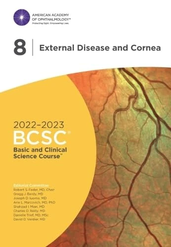
2022-2023 Basic and Clinical Science Course, Section 08: External Disease and Cornea
American Academy of Ophthalmology
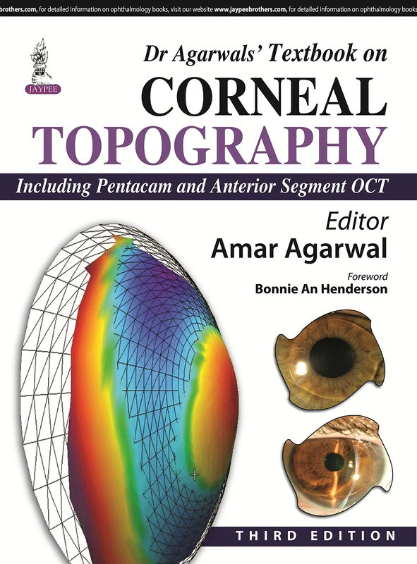
Dr Agarwal's Textbook on Corneal Topography
Jaypee Brothers Medical Publishers

2016-2017 Basic and Clinical Science Course (BCSC): Section 8: External Disease and Cornea
American Academy of Ophthalmology
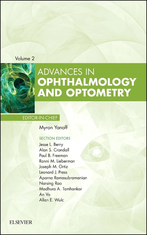
Advances in Ophthalmology and Optometry, 2017: Volume 2017 (Advances, Volume 2017)
Elsevier