We can't find the internet
Attempting to reconnect
Something went wrong
Hang in there while we get back on track
£106.60
It has never been this cheap. We have no record of a lower price.
£107 today · cheaper than every other day in the last 3 months
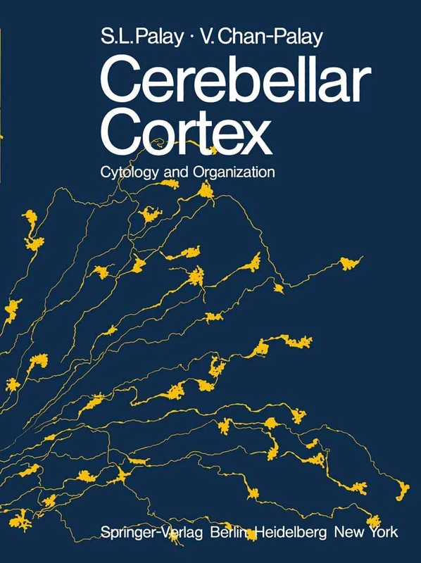
Springer Cerebellar Cortex: Cytology and Organization Book
We'll watch every seller, every day. One email when your price arrives.
Price History & Forecast
Grey patches = out of stock. Cheaper = lower on the chart. Hover for exact prices.
Last 49 days · 49 data points (no recent data)
NEW HERE?
Amazon shows you one price. We show you all of them.
Tosheroon watches Amazon prices so you don't have to. Every product on Amazon has a price history — we make it visible. Set the price you'd actually pay, and we'll email you the second it gets there. No app, no account, one email.
WHAT'S ON THIS PAGE
when this has been cheap or pricey
where the price is heading next
all-time high & low, recent range
name your number, we'll email you
Price Distribution
Price distribution over 49 days • 1 price levels
Price Analysis
Most common price: £107 (49 days, 100.0%)
Price range: £107 - £107
Price levels: 1 different prices over 49 days
Description
Key Features
This Springer publication covers the cytology and organization of the cerebellar cortex.
The text connects modern neuroanatomy to the historical work of Ramon y Cajal.
Content is based on early electron microscopic studies of the central nervous system.
Includes historical accounts of early synapse identification through electron micrographs.
Provides a detailed look at the study of cerebellar glomerulus structures.
Product Specifications
- Brand
- Springer
- Format
- paperback
- ASIN
- 3642655831
- Domain
- Amazon UK
- Release Date
- 17 November 2011
- Listed Since
- 20 September 2013
Barcode
No barcode data available
Similar Products You Might Like

The Cerebellum as a Neuronal Machine
Springer
The Cerebellum Revisited
Springer
The Cerebellum―New Vistas: 6 (Experimental Brain Research Series, 6)
Springer
The Neuronal Codes of the Cerebellum
Elsevier

Neurocytology: Fine Structure of Neurons, Nerve Processes, and Neuroglial Cells
Springer

International Review of Neurobiology (Volume 63)
Elsevier

Neuroglia
Oxford University Press

Microglia: Methods and Protocols: 2034 (Methods in Molecular Biology, 2034)
Humana

Afferent and Intrinsic Organization of Laminated Structures in the Brain: 7th International Neurobiology Meeting: 1 (Experimental Brain Research Series, 1)
Springer
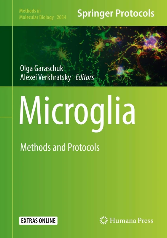
Microglia: Methods and Protocols: 2034 (Methods in Molecular Biology, 2034)
Humana

Cerebellar Learning (Volume 210) (Progress in Brain Research, Volume 210)
Elsevier

Neural Progenitor Cells: Methods and Protocols: 1059 (Methods in Molecular Biology, 1059)
Humana

Academic Press Neural Development - Vol 69 Biology Book
Elsevier
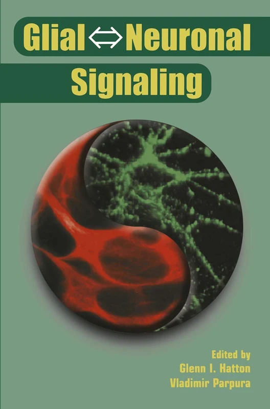
Glial ⇔ Neuronal Signaling
Springer